“X-ray” might sound like something out of a sci-fi movie, but in orthodontics, it’s a common and crucial tool that lights the way to a perfect smile. At Tilghman Orthodontics, Dr. Tilghman uses this advanced imaging technology to chart the course of each patient’s treatment precisely. These powerful glimpses beneath the gum line allow us to predict and perfect every detail of your orthodontic care. Let’s first explore the innovative X-ray technologies that make all of this possible.
Advanced X-Ray Technologies at Tilghman Orthodontics
At Tilghman Orthodontics, we employ state-of-the-art X-ray technologies that enhance our ability to diagnose accurately and plan treatments effectively. Here’s a closer look at how these technologies shape our orthodontic strategies:
- Digital X-rays: Dr. Tilghman utilizes digital X-ray systems, which are known for their low radiation levels compared to traditional X-rays. These systems produce instant, high-resolution images that are crucial for assessing treatment progress and making immediate adjustments.
- 3D Imaging: For more complex cases, 3D imaging gives a comprehensive view of the patient’s dental anatomy. This allows for a deeper analysis and understanding of the interaction between teeth, bones, and soft tissues, which is essential for crafting precise orthodontic solutions.
- Enhanced Diagnostic Capabilities: The clarity and detail provided by these advanced technologies allow for a level of diagnostic accuracy that was previously unattainable, ensuring that every aspect of the treatment plan is based on thorough and precise information.
By integrating these advanced imaging solutions, Tilghman Orthodontics not only adheres to the highest standards of orthodontic care but also ensures that each patient receives the most effective and efficient treatment possible. Next, we will explore the specific types of X-rays used at our practice and how each one contributes to the unique needs of our patients’ treatments.
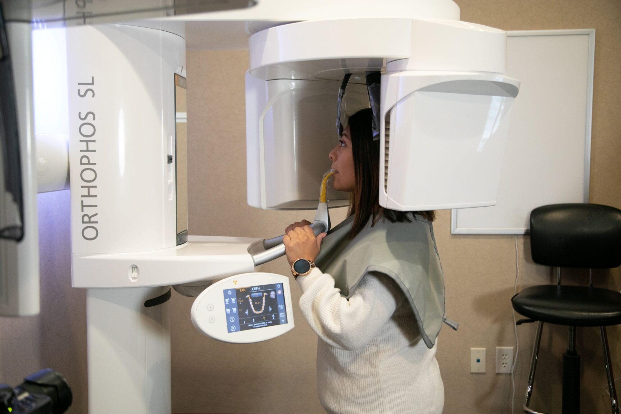
Types of Orthodontic X-Rays
We utilize a variety of X-ray types at Tilghman Orthodontics, each serving a specific purpose to enhance treatment accuracy and patient outcomes. Understanding the role of each X-ray helps in tailoring our approach to meet the precise needs of each patient’s orthodontic situation:
Panoramic X-rays
These provide a comprehensive view of the entire mouth in a single image, which is essential for assessing overall tooth alignment and jawbone health. Panoramic X-rays are particularly useful in planning the initial stages of treatment and in monitoring the eruption of teeth, especially in younger patients.
Cephalometric X-rays
Offering a side view of the head, cephalometric X-rays allow Dr. Tilghman to evaluate the teeth in relation to the jaw and the rest of the facial structure. This is crucial for planning treatments that involve adjustments to the bite or jaw alignment.
Intraoral X-rays
Focused and detailed intraoral X-rays are critical for examining specific areas of concern. They provide a close-up view of individual teeth, highlighting fine details such as the condition of tooth roots and the exact positioning of each tooth, which is vital for precise bracket placement.
Each type of X-ray plays a crucial role in our comprehensive diagnostic process, ensuring that nothing is overlooked. Up next, we’ll dive into how we use these X-rays to monitor treatment progress and make real-time adjustments, keeping each patient’s treatment on track towards achieving a perfect smile.

Monitoring Treatment Progress with X-Rays
Effective orthodontic care relies heavily on our ability to track treatment progress and respond with precision. At Tilghman Orthodontics, we harness the power of X-rays not just to visualize the starting point but to guide the entire treatment process with dynamic adjustments. Here’s how we incorporate X-ray monitoring into our patient care:
Key Milestones Tracked by X-Rays
- Initial Alignment Check: Soon after the braces are placed, X-rays help us verify that all components are positioned accurately to influence tooth movement correctly.
- Mid-Treatment Assessment: Midway through the treatment, X-rays provide a detailed look at the shifts in teeth alignment, allowing Dr. Tilghman to assess whether the treatment is progressing as planned or if any strategic modifications are needed.
- Pre-Completion Evaluation: Before concluding the treatment, a final set of X-rays ensures that all objectives have been met and that the teeth and jaw alignment are in their optimal positions.
Real-Time Adjustments Enabled by X-Ray Insights
- Adaptive Treatment Planning: Based on the latest X-ray data, Dr. Tilghman can adjust the pressure exerted by braces or recalibrate the alignment path, ensuring the treatment remains efficient and effective.
- Predictive Adjustments: Anticipating how teeth might move in the coming months allows for proactive adjustments today, minimizing potential issues and streamlining the path to the desired outcome.
Educational Benefits for Patients
- Visual Progress Sharing: During consultations, Dr. Tilghman uses X-ray images to show patients the changes in their dental structure, helping them visualize their progress and understand the treatment process.
- Engagement and Reassurance: These discussions demystify the orthodontic procedures, reinforcing patient confidence and comfort with the ongoing treatment.
Through careful monitoring and strategic adjustments guided by X-ray imaging, Tilghman Orthodontics ensures each patient’s treatment trajectory is not only adhered to but optimized for the best possible results. Next, we’ll explore how patients can prepare for X-ray sessions, ensuring they are comfortable and informed throughout their treatment journey.
Preparing for an X-Ray: What Patients Should Know
Visiting Tilghman Orthodontics for an X-ray is a straightforward and essential part of your treatment plan. Here’s how to prepare:
- Pre-Procedure: Remove any jewelry or metal accessories to prevent interference with the images. Wearing loose, comfortable clothing is also recommended.
- During the Procedure: Our team will guide you into the proper position, ensuring the best possible image quality while keeping you comfortable.
- Post-Procedure: Dr. Tilghman will review the X-rays with you, explaining the findings and discussing any necessary adjustments to your treatment plan.
This preparation ensures a smooth and efficient X-ray process, contributing to the success of your orthodontic treatment.

Clear View to a Stunning Smile
At Tilghman Orthodontics, every X-ray is a step closer to your ideal smile. Dr. Tilghman and our team expertly wield this technology to perfect your path to orthodontic success. Curious about the clarity X-rays can bring to your smile? Schedule a free consultation at our Berlin or Salisbury locations today and see the transformation for yourself!